



Key points
- Consider Cyclospora as a potential cause of prolonged diarrheal illness, particularly in patients with a history of recent travel to Cyclospora-endemic areas such as tropical and subtropical regions.
- Testing for Cyclospora is not routinely conducted in most U.S. laboratories. Healthcare providers should specifically request testing when cyclosporiasis is suspected.
Considerations
Consider Cyclospora as a potential cause of prolonged diarrheal illness, particularly in patients with a history of recent travel to Cyclospora-endemic areas such as tropical and subtropical regions. Testing for Cyclospora is not routinely conducted in most U.S. laboratories, even in stool being tested for parasites. Similarly, not all gastrointestinal polymerase chain reaction (PCR) panels include a target for Cyclospora. Healthcare providers should specifically request testing for Cyclospora if indicated.
Diagnosis
Cyclospora infection is diagnosed by examining stool specimens. Diagnosis can be difficult, in part because even symptomatic patients might not shed enough oocysts in stool to be readily detectable by laboratory examinations. Patients might need to submit several specimens collected on different days.
Testing
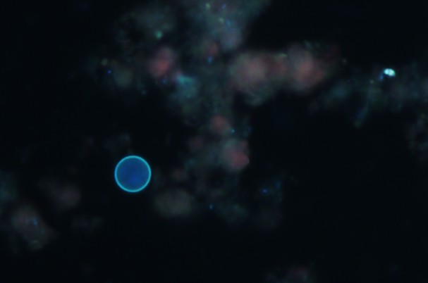
Laboratories should use sensitive recovery methods (concentration procedures) and detection methods that highlight Cyclospora oocysts. The oocysts can be stained with modified acid-fast or modified ("hot") safranin techniques. Cyclospora oocysts also are autofluorescent, meaning that when stool containing the parasite is viewed under an ultraviolet (UV) fluorescence microscope, the oocysts appear blue or green (see image above) against a black background. Molecular diagnostic methods such as PCR analysis are used to look for the parasite's DNA in the stool.
Detecting oocysts
- Cyclospora oocysts are easily overlooked; low-level shedding (~1–2 logs lower than for Cryptosporidium species) is common. To maximize recovery of Cyclospora oocysts, first concentrate the stool specimen—such as by the formalin-ethyl acetate technique (centrifuge for 10 minutes at 500 x g)—and then examine a wet mount or a stained slide (or both) of the sediment.
- Cyclospora oocysts are ~8–10 micrometers in diameter. In contrast, Cryptosporidium parvum/hominis oocysts are ~4–6 micrometers in diameter.
- Ultraviolet fluorescence microscopy (UV excitation filter set at 330–365 nm or 450–490 nm) is a sensitive technique for rapidly examining stool sediments for Cyclospora oocysts, which stand out because they autofluoresce (Cryptosporidium parvum/hominis oocysts do not). If suspect Cyclospora oocysts are found, bright-field, phase contrast, or differential interference contrast microscopy can then be used to confirm that the structures have the characteristic morphologic features of Cyclospora oocysts (i.e., nonrefractile spheres that contain undifferentiated cytoplasm or refractile globules).
- On a modified acid-fast stained slide of stool, Cyclospora oocysts typically are variably acid fast (i.e., in the same field, oocysts may be unstained or stain from light pink to deep red or purple). Unstained oocysts characteristically have a wrinkled (hyaline) appearance.
- If a "hot" modified safranin technique is used, Cyclospora oocysts uniformly stain a brilliant reddish orange.
This page was last updated on this date. Updates may include minor edits, image changes, or other modifications to page content.
The information on this page was last reviewed by subject matter experts to ensure accuracy.